Subscribe

RSS Feed (xml)

Powered By

Skin Design:
Free Blogger Skins

Powered by Blogger

Showing posts with label Cell Biology. Show all posts

Thursday, April 30, 2009

article : What Is Cell Biology?


All forms of life are made from these tiny units ... cells, and there are only two main types; prokaryotes and eukaryotes. Explore the following topics and learn more about cells and the cellular processes that that are the common denominator shared by all living things.

SPO Cell Biology Topics

Prokaryotic and Eukaryotic Cells

Prokaryotes, the simplest form of life, are evolutionarily ancient, and for billions of years the only form of life. Now the domains Archea and Eubacteria represent the extant (currently living) types of prokaryotic cells.


While prokaryotic cells include bacteria and bacteria-like Archea, eukaryotic cells are... everything else. From the cells that make up your body to the tiny blob-like amoeba, most living things that we are familiar with are eukaryotes. Eukaryotic cells are distinguished from prokaryotes by the presence of a nucleus and other membrane-bound organelles. The following articles provide information specific to eukaryotes.

Chemistry of the Cell

Think that chemistry has nothing to do with biology? Think again. In order to understand cells and cellular processes, you must have at least a very basic understanding of inorganic and organic chemistry.Click on this topic link to access the articles that will bring you up to speed.


Cellular Metabolism

We are all familiar with the term 'metabolism', but what does it really mean? The following articles explain how the cells of our bodies (and cells of all living things) turn food energy into ATP energy that can be used to fuel cellular reactions.

Cell Division - Mitosis and Meiosis

Cell division is required for living things to grow, develop and reproduce. Mitosis produces cells that are clones, identical to the parent cell. Meiosis is the form of cell division that produced gametes (sperm and eggs). See the links below for more detailed information on cell division.


Molecular Genetics

Learn about nucleic acids, the organic molecules that contain and transmit the genetic code to build the vast array of proteins that make you truly unique.


Stem Cells

What are stem cells? How are they collected, stored, grown and transplanted?

This link will take you to an article series that should answer some of your questions regarding embryonic and adult stem cells, umbilical cord blood banking and more.

Virtual Cell Biology Classroom

Materials used to supplement an introductory college-level Cell Biology Course. The Virtual Cell Biology Classroom provides access to a wide range of educational resources including Power Point Lectures, Study Guides, Review Questions and Practice Test Questions

Sources

Images

Epithilial Cells: WikiBooks Cell Biology Text

Cell Endomembrane System: Mariana Ruiz

Macrophage: National Science Council


Read More......

Thursday, April 10, 2008

article : Cell Division

Cell division is a process by which a cell, called the parent cell, divides into two cells, called daughter cells. Cell division is usually a small segment of a larger cell cycle. In meiosis however, a cell is permanently transformed and cannot divide again.

For simple unicellular organisms such as the Amoeba, one cell division reproduces an entire organism. On a larger scale, cell division can create progeny from multicellular organisms, such as plants that grow from cuttings. Cell division also enables sexually reproducing organisms to develop from the one-celled zygote, which itself was produced by cell division from gametes. And after growth, cell division allows for continual renewal and repair of the organism.[1] A human being's body experiences about 10,000 trillion cell divisions in a lifetime.[2]

The primary concern of cell division is the maintenance of the original cell's genome. Before division can occur, the genomic information which is stored in chromosomes must be replicated, and the duplicated genome separated cleanly between cells. A great deal of cellular infrastructure is involved in keeping genomic information consistent between "generations".

Variants

Three types of cell division

Cells are classified into two categories: simple, non-nucleated prokaryotic cells, and complex, nucleated eukaryotic cells. By dint of their structural differences, eukaryotic and prokaryotic cells do not divide in the same way.

Furthermore, the pattern of cell division that transforms eukaryotic stem cells into gametes (sperm in males or ova in females) is different from that of eukaryotic somatic (non-germ) cells.

Prokaryotic cells

Main article: binary fission

Prokaryotic cells are generally much more simple in structure when compared to eukaryotic cells. They contain non-membranous organelles, lack a cell nucleus, and have a simplistic genome: only one circular chromosome of limited size. Therefore, prokaryotic cell division, a process known as binary fission, is fast. The chromosome is duplicated prior to division. The two copies of the chromosome attach to opposing sides of the cellular membrane. Cytokinesis, the physical separation of the cell, occurs immediately.

Eukaryotic cells

Main article: mitosis
  • Mitosis: The division of the nucleus, separating the duplicated genome into two sets identical to the parent's.
  • Cytokinesis: The division of the cytoplasm, separating the organelles and other cellular components.
  • Meiosis: The division of the nucleus in sex cells, making one cell into four sex cells identical to the parent sex cell.

Degradation

Multicellular organisms replace worn-out cells through cell division. In some animals, however, cell division eventually halts. In humans this occurs on average, after 52 divisions, known as the Hayflick limit. The cell is then referred to as senescent. Senescent cells deteriorate and die, causing the body to age. Cells stop dividing because the telomeres, protective bits of DNA on the end of a chromosome, become shorter with each division and eventually can no longer protect the chromosome. Cancer cells, on the other hand, are immortal. An enzyme called telomerase, present in large quantites in cancerous cells, rebuilds the telomeres, allowing division to continue indefinitely.

See also

References

  1. Morgan DO. (2007) "The Cell Cycle: Principles of Control" London: New Science Press.
  2. J.M.Turner Fetus into Man (1978, 1989). Harvard University Press. ISBN 0-674-30692-9
http://en.wikipedia.

Read More......

article ; Mitosis in Cell (2)

mītōˈsĭs, mĭ–, process of nuclear division in a living cell by which the carriers of hereditary information, or the chromosomes, are exactly replicated and the two copies distributed to identical daughter nuclei. Mitosis is almost always accompanied by cell division (cytokinesis), and the latter is sometimes considered a part of the mitotic process. The pattern of mitosis is fundamentally the same in all cells. However, while animal cells apparently divide by pinching into two separate cells, plant cells develop a cell plate, which becomes a cellulose cell wall between the two daughter cells. The importance of mitosis is the maintenance of the chromosomal set; each cell formed receives chromosomes that are alike in composition and equal in number to the chromosomes of the parent cell.

The Stages of Mitosis

Mitosis is simply described as having four stages—prophase, metaphase, anaphase, and telophase; the steps follow one another without interruption. The entire four-stage division process averages about one hour in duration, and the period between cell divisions, called interphase or interkinesis, varies greatly but is considerably longer.

During interphase the chromosomes are dispersed in the nucleus and appear as a network of long, thin threads or filaments, called the chromatin. At some point before prophase begins, the chromosomes replicate themselves to form pairs of identical sister chromosomes, or chromatids; the deoxyribose nucleic acid (DNA) of the chromosomes is synthesized only during interphase, not while mitosis is in process.

During prophase the two chromatids remain attached to one another at a region called the centromere, but each contracts into a compact tightly coiled body; the nucleolus and, in most cases, the nuclear envelope break down and disappear. Also during prophase the spindle begins to form. In animal cells the centrioles separate and move apart, and radiating bundles of fibers, called asters, appear around them. Some sets of fiber run from one centriole to the other; these are the spindle fibers. In plant cells the spindle forms without centrioles.

During metaphase the chromosomes congregate at a plane midway between the two ends to which the spindle tapers. This is called the equatorial plane and marks the point where the whole cell will divide when nuclear division is completed; the ends of the spindle are the poles to which the chromatids will migrate. The chromatids are attached to the spindle fibers at the centromeres.

During anaphase the two chromatids of each chromosome separate and move to opposite poles, as if pulled along the spindle fibers by the centromeres. During telophase new nuclear envelopes form around the two groups of daughter chromosomes (as they are now called), the new nucleoli begin to appear, and eventually, as the formation of the two daughter nuclei is completed, the spindle fibers disappear. The chromosomes uncoil to assume their dispersed distribution within the interphased nucleus. Cytokinesis, which may begin before or after mitosis is completed, finally separates the daughter nuclei into two new individual daughter cells.

A considerable variance in the degree and timing of these stages exists across species, and cells can be classified by their mitotic characteristics. Despite the relative ease of observation of the physical stages of mitosis under the microscope (primarily because the chromosomes stain readily when in their coiled state), the exact chemical and kinetic nature of mitosis is not yet fully understood. For instance, the spindle has been determined to consist largely of thin, elongate tubules called microtubules, but their functions have yet to be understood.

Meiosis and Amitosis

Mitotic division is the method of nuclear division of the somatic (body) cells, as distinguished from the gametes, or sex cells (eggs and sperm). In sexual reproduction, i.e., by the union of two gametes, the complex process of meiosis takes place, which produces cells that each contain only half the normal number of chromosomes. Direct cell division, in which the nucleus simply cleaves in two (sometimes but not always followed by division of the cytoplasm), is called amitosis and is very rare.

____________________

The Columbia Encyclopedia, Sixth Edition Copyright© 2004, Columbia University Press. Licensed from Lernout & Hauspie Speech Products N.V. All rights reserved.

Read More......

article : Mitosis in Cell (1)

Jump to: navigation, search
Mitosis divides the chromosomes in a cell nucleus.

Mitosis is the process by which a cell duplicates the chromosomes in its cell nucleus, in order to generate two, identical, daughter nuclei. [1]. It is generally followed immediately by cytokinesis, which divides the nuclei, cytoplasm, organelles and cell membrane into two daughter cells containing roughly equal shares of these cellular components. Mitosis and cytokinesis together define the mitotic (M) phase of the cell cycle, the division of the mother cell into two daughter cells, each with the genetic equivalent of the parent cell.

Mitosis occurs exclusively in eukaryotic cells, but occurs in different ways in different species. For example, animals undergo an "open" mitosis, where the nuclear envelope breaks down before the chromosomes separate, while fungi such as Aspergillus nidulans and Saccharomyces cerevisiae (yeast) undergo a "closed" mitosis, where chromosomes divide within an intact cell nucleus.[2] Prokaryotic cells, which lack a nucleus, divide by a process called binary fission.

The process of mitosis is complex and highly regulated. The sequence of events is divided into phases, corresponding to the completion of one set of activities and the start of the next. These stages are prophase, prometaphase, metaphase, anaphase and telophase. During the process of mitosis the pairs of chromosomes condense and attach to fibers that pull the sister chromatids to opposite sides of the cell. The cell then divides in cytokinesis, to produce two identical daughter cells.[3]

Because cytokinesis usually occurs in conjunction with mitosis, "mitosis" is often used interchangeably with "mitotic phase". However, there are many cells where mitosis and cytokinesis occur separately, forming single cells with multiple nuclei. This occurs most notably among the fungi and slime moulds, but is found in various different groups. Even in animals, cytokinesis and mitosis may occur independently, for instance during certain stages of fruit fly embryonic development.[4] Errors in mitosis can either kill a cell through apoptosis or cause mutations that may lead to cancer.

Overview

The primary result of mitosis is the division of the parent cell's genome into two daughter cells. The genome is composed of a number of chromosomes, complexes of tightly-coiled DNA that contain genetic information vital for proper cell function. Because each resultant daughter cell should be genetically identical to the parent cell, the parent cell must make a copy of each chromosome before mitosis. This occurs during S phase, in interphase, the period that precedes the mitotic phase in the cell cycle where preparation for mitosis occurs.[5]

Each new chromosome now contains two identical copies of itself, called sister chromatids, attached together in a specialized region of the chromosome known as the centromere. Each sister chromatid is not considered a chromosome in itself, and a chromosome does not always contain two sister chromatids.

In most eukaryotes, the nuclear envelope that separates the DNA from the cytoplasm disassembles. The chromosomes align themselves in a line spanning the cell. Microtubules, essentially miniature strings, splay out from opposite ends of the cell and shorten, pulling apart the sister chromatids of each chromosome.[6] As a matter of convention, each sister chromatid is now considered a chromosome, so they are renamed to sister chromosomes. As the cell elongates, corresponding sister chromosomes are pulled toward opposite ends. A new nuclear envelope forms around the separated sister chromosomes.

As mitosis completes cytokinesis is well underway. In animal cells, the cell pinches inward where the imaginary line used to be, (the pinching of the cell membrane to form the two daughter cells is called cleavage furrow) separating the two developing nuclei. In plant cells, the daughter cells will construct a new dividing cell wall between each other. Eventually, the mother cell will be split in half, giving rise to two daughter cells, each with an equivalent and complete copy of the original genome.

Prokaryotic cells undergo a process similar to mitosis called binary fission. However, prokaryotes cannot be properly said to undergo mitosis because they lack a nucleus and only have a single chromosome with no centromere.[7]

Phases

Interphase

The cell cycle
The cell cycle


The mitotic phase is a relatively short period of the cell cycle. It alternates with the much longer interphase, where the cell prepares itself for cell division. Interphase is divided into three phases, G1 (first gap), S (synthesis), and G2 (second gap). During all three phases, the cell grows by producing proteins and cytoplasmic organelles. However, chromosomes are replicated only during the S phase. Thus, a cell grows (G1), continues to grow as it duplicates its chromosomes (S), grows more and prepares for mitosis (G2), and divides (M).[5]

Preprophase

Main article: Preprophase

In plant cells only, prophase is preceded by a pre-prophase stage. In highly vacuolated plant cells, the nucleus has to migrate into the center of the cell before mitosis can begin. This is achieved through the formation of a phragmosome, a transverse sheet of cytoplasm that bisects the cell along the future plane of cell division. In addition to phragmosome formation, preprophase is characterized by the formation of a ring of microtubules and actin filaments (called preprophase band) underneath the plasmamembrane around the equatorial plane of the future mitotic spindle and predicting the position of cell plate fusion during telophase. The cells of higher plants (such as the flowering plants) lack centrioles. Instead, spindle microtubules aggregate on the surface of the nuclear envelope during prophase. The preprophase band disappears during nuclear envelope disassembly and spindle formation in prometaphase.[8]

Prophase

Prophase: The two round objects above the nucleus are the centrosomes. Note the condensed chromatin.
Main article: Prophase

Normally, the genetic material in the nucleus is in a loosely bundled coil called chromatin. At the onset of prophase, chromatin condenses together into a highly ordered structure called a chromosome. Since the genetic material has already been duplicated earlier in S phase, the replicated chromosomes have two sister chromatids, bound together at the centromere by the cohesion complex. Chromosomes are visible at high magnification through a light microscope.

Close to the nucleus are two centrosomes. Each centrosome, which was replicated earlier independent of mitosis, acts as a coordinating center for the cell's microtubules. The two centrosomes nucleate microtubules (which may be thought of as cellular ropes or poles) by polymerizing soluble tubulin present in the cytoplasm. Molecular motor proteins create repulsive forces that will push the centrosomes to opposite side of the nucleus. The centrosomes are only present in animals. In plants the microtubules form independently.

Some centrosomes contain a pair of centrioles that may help organize microtubule assembly, but they are not essential to formation of the mitotic spindle.[9]

Prometaphase

Prometaphase: The nuclear membrane has degraded, and microtubules have invaded the nuclear space. These microtubules can attach to kinetochores or they can interact with opposing microtubules.
Main article: Prometaphase

The nuclear envelope disassembles and microtubules invade the nuclear space. This is called open mitosis, and it occurs in most multicellular organisms. Fungi and some protists, such as algae or trichomonads, undergo a variation called closed mitosis where the spindle forms inside the nucleus or its microtubules are able to penetrate an intact nuclear envelope.[10][11]

Each chromosome forms two kinetochores at the centromere, one attached at each chromatid. A kinetochore is a complex protein structure that is analogous to a ring for the microtubule hook; it is the point where microtubules attach themselves to the chromosome.[12] Although the kinetochore structure and function are not fully understood, it is known that it contains some form of molecular motor.[13] When a microtubule connects with the kinetochore, the motor activates, using energy from ATP to "crawl" up the tube toward the originating centrosome. This motor activity, coupled with polymerisation and depolymerisation of microtubules, provides the pulling force necessary to later separate the chromosome's two chromatids.[13]

When the spindle grows to sufficient length, kinetochore microtubules begin searching for kinetochores to attach to. A number of nonkinetochore microtubules find and interact with corresponding nonkinetochore microtubules from the opposite centrosome to form the mitotic spindle.[14] Prometaphase is sometimes considered part of prophase.

Metaphase

Metaphase: The chromosomes have aligned at the metaphase plate.
Main article: Metaphase

As microtubules find and attach to kinetochores in prometaphase, the centromeres of the chromosomes convene along the metaphase plate or equatorial plane, an imaginary line that is equidistant from the two centrosome poles.[14] This even alignment is due to the counterbalance of the pulling powers generated by the opposing kinetochores, analogous to a tug-of-war between equally strong people. In certain types of cells, chromosomes do not line up at the metaphase plate and instead move back and forth between the poles randomly, only roughly lining up along the midline. Metaphase comes from the Greek μετα meaning "after."

Because proper chromosome separation requires that every kinetochore be attached to a bundle of microtubules (spindle fibers) , it is thought that unattached kinetochores generate a signal to prevent premature progression to anaphase[1] without all chromosomes being aligned. The signal creates the mitotic spindle checkpoint.[15]

Anaphase

Early anaphase: Kinetochore microtubules shorten
Main article: Anaphase

When every kinetochore is attached to a cluster of microtubules and the chromosomes have lined up along the metaphase plate, the cell proceeds to anaphase (from the Greek ανα meaning “up,” “against,” “back,” or “re-”).

Two events then occur; First, the proteins that bind sister chromatids together are cleaved, allowing them to separate. These sister chromatids turned sister chromosomes are pulled apart by shortening kinetochore microtubules and move toward the respective centrosomes to which they are attached. Next, the nonkinetochore microtubules elongate, pushing the centrosomes (and the set of chromosomes to which they are attached) apart to opposite ends of the cell. The force that causes the centrosomes to move towards the ends of the cell is still unknown, although there is a theory that suggests that the rapid assembly and and breakdown of microtubules may cause this movement.[16]

These two stages are sometimes called early and late anaphase. Early anaphase is usually defined as the separation of the sister chromatids, while late anaphase is the elongation of the microtubules and the microtubules being pulled farther apart. At the end of anaphase, the cell has succeeded in separating identical copies of the genetic material into two distinct populations.

Telophase

Telophase: The decondensing chromosomes are surrounded by nuclear membranes. Note cytokinesis  has already begun, the pinching is known as the cleavage furrow.
Telophase: The decondensing chromosomes are surrounded by nuclear membranes. Note cytokinesis has already begun, the pinching is known as the cleavage furrow.
Main article: Telophase

Telophase (from the Greek τελος meaning "end") is a reversal of prophase and prometaphase events. It "cleans up" the after effects of mitosis. At telophase, the nonkinetochore microtubules continue to lengthen, elongating the cell even more. Corresponding sister chromosomes attach at opposite ends of the cell. A new nuclear envelope, using fragments of the parent cell's nuclear membrane, forms around each set of separated sister chromosomes. Both sets of chromosomes, now surrounded by new nuclei, unfold back into chromatin. Mitosis is complete, but cell division is not yet complete.

Cytokinesis

Main article: Cytokinesis

Cytokinesis is often mistakenly thought to be the final part of telophase, however cytokinesis is a separate process that begins at the same time as telophase. Cytokinesis is technically not even a phase of mitosis, but rather a separate process, necessary for completing cell division. In animal cells, a cleavage furrow (pinch) containing a contractile ring develops where the metaphase plate used to be, pinching off the separated nuclei.[17] In both animal and plant cells, cell division is also driven by vesicles derived from the Golgi apparatus, which move along microtubules to the middle of the cell. [18] In plants this structure coalesces into a cell plate at the center of the phragmoplast and develops into a cell wall, separating the two nuclei. The phragmoplast is a microtubule structure typical for higher plants, whereas some green algae use a phycoplast microtubule array during cytokinesis.[19] Each daughter cell has a complete copy of the genome of its parent cell. The end of cytokinesis marks the end of the M-phase.

Significance

The importance of mitosis is the maintenance of the chromosomal set; each cell formed receives chromosomes that are alike in composition and equal in number to the chromosomes of the parent cell. Transcription is generally believed to cease during mitosis, but epigenetic mechanisms such as bookmarking function during this stage of the cell cycle to ensure that the "memory" of which genes were active prior to entry into mitosis are transmitted to the daughter cells.[20]

Consequences of errors

Although errors in mitosis are rare, the process may go wrong, especially during early cellular divisions in the zygote. Mitotic errors can be especially dangerous to the organism because future offspring from this parent cell will carry the same disorder.

In non-disjunction, a chromosome may fail to separate during anaphase. One daughter cell will receive both sister chromosomes and the other will receive none. This results in the former cell having three chromosomes coding for the same thing (two sisters and a homologue), a condition known as trisomy, and the latter cell having only one chromosome (the homologous chromosome), a condition known as monosomy. These cells are considered aneuploidic cells and these abnormal cells can cause cancer.[21]

Mitosis is a traumatic process. The cell goes through dramatic changes in ultrastructure, its organelles disintegrate and reform in a matter of hours, and chromosomes are jostled constantly by probing microtubules. Occasionally, chromosomes may become damaged. An arm of the chromosome may be broken and the fragment lost, causing deletion. The fragment may incorrectly reattach to another, non-homologous chromosome, causing translocation. It may reattach to the original chromosome, but in reverse orientation, causing inversion. Or, it may be treated erroneously as a separate chromosome, causing chromosomal duplication. The effect of these genetic abnormalities depend on the specific nature of the error. It may range from no noticeable effect, cancer induction, or organism death.

Endomitosis

Endomitosis is a variant of mitosis without nuclear or cellular division, resulting in cells with many copies of the same chromosome occupying a single nucleus. This process may also be referred to as endoreduplication and the cells as endoploid.[4] An example of a cell that goes through endomitosis is the megakaryocyte.[22]

Timeline in pictures

Real mitotic cells can be visualized through the microscope by staining them with fluorescent antibodies and dyes. These light micrographs are included below.

http//wikipedia.org

Read More......

article : ANIMAL CELLS AND TISSUES

By Michael J. Farabee, Ph.D., Estrella Mountain Community College, Update 12/06

Table of Contents

Organization of the Animal Body | Epithelial Tissue | Connective Tissue | Muscle Tissue

Nervous Tissue | Learning Objectives | Terms | Review Questions | Links

Organization of the Animal Body

Animals are multicellular heterotrophs whose cells lack cell walls. At some point during their lives, all animals are capable of movement, although not all animals have muscles they use for this. In the most commonly encountered animals, the mobile stage is the adult, although some animals (such as corals and sponges) have sessile (or nonmobile) adult phases and mobile juvenile forms. Both animal and plant evolutionary history show the development of multicellularity and the move from water to land (as well as a secondary adaptation back to water, for example dolphins, whales, duckweed, and elodea).

Animals developed external or internal skeletons to provide support, skin to prevent or lessen water loss, muscles that allowed them to move in search of food, brains and nervous systems for integration of stimuli, and internal digestive systems.

Organs in animals are composed of a number of different tissue types. For example, the stomach shown in Figure 1, has epithelial tissue making linings and secreting gastric juices, connective tissues

Figure 1. Cells and tissues that comprise the stomach. Image from Purves et al., Life: The Science of Biology, 4th Edition, by Sinauer Associates (www.sinauer.com) and WH Freeman (www.whfreeman.com), used with permission.

Plants are simpler organisms than animals, having three organ systems and fewer organs than do vertebrate animals. Organs are composed of tissues, which are in turn composed of cells. Plants have three tissue types: ground, dermal, and vascular. Animals have four: epithelial, connective, muscle, and bone.

Epithelial Tissue | Back to Top

Epithelial tissue covers body surfaces and lines body cavities. Functions include lining, protecting, and forming glands. Three types of epithelium occur:

  • Squamous epithelium is flattened cells.
  • Cuboidal epithelium is cube-shaped cells.
  • Columnar epithelium consists of elongated cells.

Any epithelium can be simple or stratified. Simple epithelium has only a single cell layer. Stratified epithelium has more than one layer of cells. Pseudostratified epithelium is a single layer of cells so shaped that they appear at first glance to form two layers.

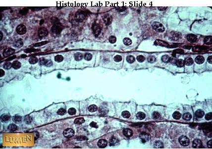
Figure 2. Cuboidal epithelium. The image is cropped from Loyola University's LUMEN site at http://www.meddean.luc.edu/lumen/MedEd/Histo/HistoImages/hl1-04.jpg. Note the single layer of simple cuboidal epithelium lining either side of a tubule.

Figure 3. Epithelium lining the intestine of a rat, as seen with SEM. This image is from http://130.102.208.100/FMRes/FMPro?-db=images.fp3&key=32816&-img, used by permission of Nanoworld.

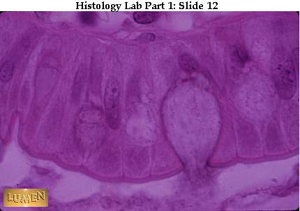
Figure 4. Columnar epithelial cells. The above image is cropped and modified from Loyola University's LUMEN site at http://www.meddean.luc.edu/lumen/MedEd/Histo/HistoImages/hl1-12.jpg. Note: I have outlined one of the columnar epithelium cells.

Functions of epithelial cells include:

  • movement materials in, out, or around the body.
  • protection of the internal environment against the external environment.
  • Secretion of a product.

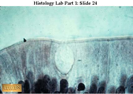
Glands can be single epithelial cells, such as the goblet cells that line the intestine. Multicellular glands include the endocrine glands. Many animals have their skin composed of epithelium. Vertebrates have keratin in their skin cells to reduce water loss. Many other animals secrete mucus or other materials from their skin, such as earthworms do.

Figure 5. Glandular epithelium. The image is from Loyola University's LUMEN site at http://www.meddean.luc.edu/lumen/MedEd/Histo/HistoImages/hl1-24.jpg).

Connective Tissue | Back to Top

Connective tissue serves many purposes in the body:

  • binding
  • supporting
  • protecting
  • forming blood
  • storing fats
  • filling space

Connective cells are separated from one another by a non-cellular matrix. The matrix may be solid (as in bone), soft (as in loose connective tissue), or liquid (as in blood). Two types of connective tissue are Loose Connective Tissue (LCT) and Fibrous Connective Tissue (FCT). Fibroblasts (LCT) are separated by a collagen fiber-containing matrix. Collagen fibers provide elasticity and flexibility. LCT occurs beneath epithelium in skin and many internal organs, such as lungs, arteries and the urinary bladder. This tissue type also forms a protective layer over muscle, nerves, and blood vessels.

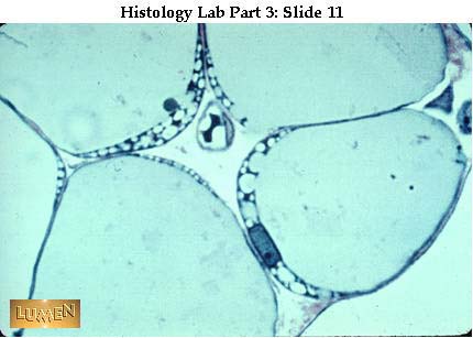
Figure 6. Adipose tissue, a type of connective tissue. The image is cropped from Loyola University's LUMEN page at http://www.meddean.luc.edu/lumen/MedEd/Histo/HistoImages/hl2-11.jpg.

Adipose tissue, shown in Figure 6, has enlarged fibroblasts storing fats and reduced intracellular matrix. Adipose tissue facilitates energy storage and insulation.

Fibrous Connective Tissue has many fibers of collagen closely packed together. FCT occurs in tendons, which connect muscle to bone. Ligaments are also composed of FCT and connect bone to bone at a joint.

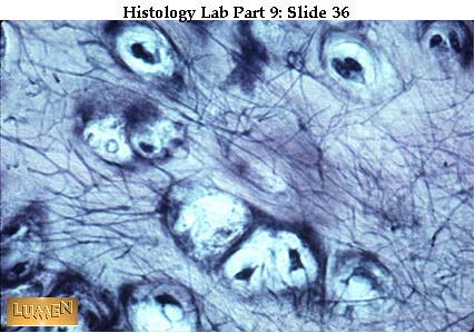
Cartilage and bone are "rigid" connective tissues. Cartilage, shown in Figure 7, has structural proteins deposited in the matrix between cells. Cartilage is the softer of the two "rigid" connective tissues. Cartilage forms the embryonic skeleton of vertebrates and the adult skeleton of sharks and rays. It also occurs in the human body in the ears, tip of the nose, and at joints such as the knee and between bones of the spinal column.

Figure 7. Cartilage, a type of "soft" connective tissue. The image is cropped from Loyola University's LUMEN page at http://www.meddean.luc.edu/lumen/MedEd/Histo/HistoImages/hl4A-36.jpg.

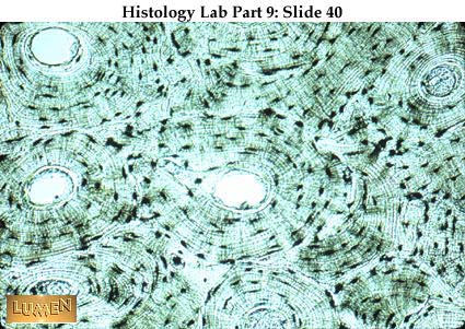
Bone, shown in Figure 8, has calcium salts in the matrix, giving it greater rigidity and strength. Bone also serves as a reservoir (or sink) for calcium. Protein fibers provide elasticity while minerals provide elasticity. Two types of bone occur. Dense bone has osteocytes (bone cells) located in lacunae connected by canaliculi. Lacunae are commonly referred to as Haversian canals. Spongy bone occurs at the ends of bones and has bony bars and plates separated by irregular spaces. The solid portions of spongy bone pick up stress.

Figure 8. Bone. The first image of bone is cropped from Loyola University's LUMEN page at http://www.meddean.luc.edu/lumen/MedEd/Histo/HistoImages/hl4A-40.jpg. Note the haversian canal and surrounded by osteocytes and a mineralized matrix. The second image shows the structure and vascularization of bone. Image from Purves et al., Life: The Science of Biology, 4th Edition, by Sinauer Associates (www.sinauer.com) and WH Freeman (www.whfreeman.com), used with permission.

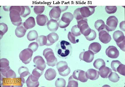
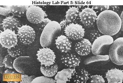
Blood is a connective tissue of cells separated by a liquid (plasma) matrix. Illustrations of blood cells are shown in Figure 9. Two types of cells occur. Red blood cells (erythrocytes) carry oxygen. White blood cells (leukocytes) function in the immune system. Plasma transports dissolved glucose, wastes, carbon dioxide and hormones, as well as regulating the water balance for the blood cells. Platelets are cell fragments that function in blood clotting.

Figure 9. Elements of the blood. The left image below is cropped from Loyola University's LUMEN site at http://www.meddean.luc.edu/lumen/MedEd/Histo/HistoImages/hl2B-51.jpg. Note the red blood cells and the single neutrophil. The right image below is cropped from Loyola University's LUMEN site at http://www.meddean.luc.edu/lumen/MedEd/Histo/HistoImages/hl2B-64.jpg. Erythrocytes as seem with the SEM. The bottom image is human red blood cells, platelets and T-lymphocyte (erythrocytes = red; platelets = yellow; T-lymphocyte = light green) (SEM x 9,900). This image is copyright Dennis Kunkel at www.DennisKunkel.com, used with permission.

Muscle Tissue | Back to Top

Muscle tissue facilitates movement of the animal by contraction of individual muscle cells (referred to as muscle fibers). Three types of muscle fibers occur in animals (the only taxonomic kingdom to have muscle cells):

  • skeletal (striated)
  • smooth
  • cardiac

Muscle tissue and organization is shown in Figure 10.

Figure 10. Organization of muscle tissue. Images from Purves et al., Life: The Science of Biology, 4th Edition, by Sinauer Associates (www.sinauer.com) and WH Freeman (www.whfreeman.com), used with permission.

Muscle fibers are multinucleated, with the nuclei located just under the plasma membrane. Most of the cell is occupied by striated, thread-like myofibrils. Within each myofibril there are dense Z lines. A sarcomere (or muscle functional unit) extends from Z line to Z line. Each sarcomere has thick and thin filaments. The thick filaments are made of myosin and occupy the center of each sarcomere. Thin filaments are made of actin and anchor to the Z line.

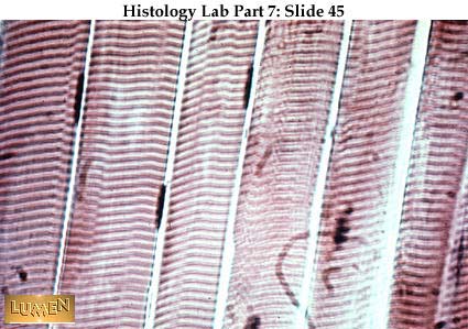
Skeletal (striated) muscle fibers, shown in Figure 11, have alternating bands perpendicular to the long axis of the cell. These cells function in conjunction with the skeletal system for voluntary muscle movements. The bands are areas of actin and myosin deposition in the cells.

Figure 11. Striated muscle cells. The left image of striated muscle fibers is cropped from Loyola University's LUMEN site at http://www.meddean.luc.edu/lumen/MedEd/Histo/HistoImages/hl3A-45.jpg. The right image is from http://130.102.208.100/FMRes/FMPro?-db=images.fp3&key=32948&-img.

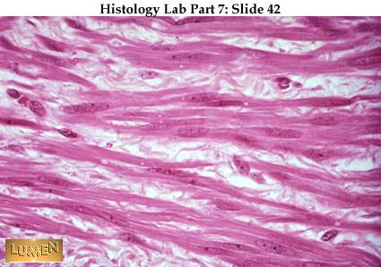
Smooth muscle fibers, shown in Figure 12, lack the banding, although actin and myosin still occur. These cells function in involuntary movements and/or autonomic responses (such as breathing, secretion, ejaculation, birth, and certain reflexes). Smooth muscle fibers are spindle shaped cells that form masses. These fibers are components of structures in the digestive system, reproductive tract, and blood vessels.

Figure 12. Smooth muscle cells. The image of smooth muscle cells is cropped from Loyola University's LUMEN site at http://www.meddean.luc.edu/lumen/MedEd/Histo/HistoImages/hl3A-42.jpg.

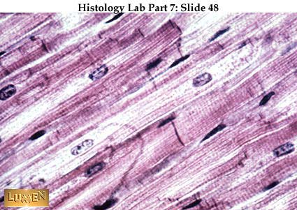
Cardiac muscle fibers are a type of striated muscle found only in the heart. The cell has a bifurcated (or forked) shape, usually with the nucleus near the center of the cell. The cells are usually connected to each other by intercalated disks, as shown in Figure 13.

Figure 13. Cardiac muscle cells. The top image of cardiac muscle cells is cropped from Loyola University's LUMEN site at http://www.meddean.luc.edu/lumen/MedEd/Histo/HistoImages/hl3A-48.jpg. Note the dark band of the intercalated disk that separates two muscle cells. The bottom image is of a heart muscle cell (nucleus, mitochondria, actin-myosin) (TEM x15,400). This image is copyright Dennis Kunkel at www.DennisKunkel.com, used with permission.

Nervous Tissue | Back to Top

Nervous tissue, shown in Figure 14, functions in the integration of stimulus and control of response to that stimulus. Nerve cells are called neurons. Each neuron has a cell body, an axon, and many dendrites. Nervous tissue is composed of two main cell types: neurons and glial cells. Neurons transmit nerve messages. Glial cells are in direct contact with neurons and often surround them.

Figure 14. Organization of a neutron. Image from Purves et al., Life: The Science of Biology, 4th Edition, by Sinauer Associates (www.sinauer.com) and WH Freeman (www.whfreeman.com), used with permission.

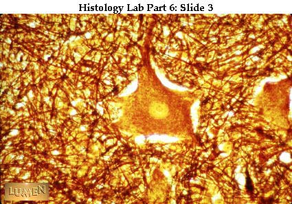
The neuron is the functional unit of the nervous system. Humans have about 100 billion neurons in their brain alone! While variable in size and shape, all neurons have three parts. Dendrites receive information from another cell and transmit the message to the cell body. The cell body contains the nucleus, mitochondria and other organelles typical of eukaryotic cells. The axon conducts messages away from the cell body. Neurons are shown in Figure 15.

Figure 15. Neurons. The left image of large multipolar neuron (center of image) is cropped from Loyola University's LUMEN site at http://www.meddean.luc.edu/lumen/MedEd/Histo/HistoImages/hl3-03.jpg. The right image shows Pyramidal Neurons from the Central Nervous System (SEM x3,960). This image is copyright Dennis Kunkel at www.DennisKunkel.com, used with permission.


http://www.emc.maricopa.edu

Read More......

article : Animal Tissue Culture and Hybridoma Technology

Animal Tissue Culture and Hybridoma Technolog - The term tissue culture refers to the culture of whole organs, tissue fragments as well as dispersed cells on a suitable nutrient medium. It can be divided into

(1) organ culture and

(2) cell culture mainly on the basis of whether the tissue organisation is retained or not.

In organ cultures, whole embryonic organs or small tissue fragments are cultured in vitro in such a manner that they retain their tissue architecture. In contrast, cell cultures are obtained either by enzymatic or mechanical dispersal of tissues into individual cells or by spontaneous migration of cells from explants; they are maintained as attached monolayers or as cell suspensions.

Freshly isolated cell cultures are called primary cultures; they are usually heterogeneous and slow growing, but are more representative of the tissue of their origin both in cell type and properties. Once a primary culture is subcultured, it gives rise to cell lines, which may either die after several subcultures (such cell lines are known as finite cell lines) or may continue to grow indefinitely (these are called continuous cell lines).

Usually, normal tissues give rise to finite cell lines, while tumours give rise to continuous cell lines. But there are several examples of continuous cell lines, which were derived from normal tissues and are themselves nontumorigenic, e.g., MDCK dog kidney, 3T3 fibroblasts, etc.

The evolution of continuous cell lines from primary cultures is supposed to involve a mutation, which alters their properties as compared to those of finite lines.

http://www.molecular-plant-biotechnology.info


Read More......

article : Animal tissue review

A review of the structure of animal tissue (connective, nervous, epithelial, and muscle) and how it is directly related to its function.

The structure of animal tissue is directly related to its function. Animals have groups of cells in the tissue, which are combined together to perform as an organ. Tissues are groups of cells with a basic structure and function. There are four major types of tissues: epithelial tissue, connective tissue, nervous tissue, and muscle tissue. Tissues are woven together with a sticky extra cellular matrix.

Epithelial tissue is a tissue that is made up of tightly packed cells that line organs and body cavities. The cells contained in the epithelium are closely joined without much material between them. Inside the epithelia the cells are locked together by tight junctions. The reasons for the tightly packed cells are to act as a barrier against mechanical injury, invading microorganisms, and fluid loss. The base of the barrier cells are attached to the basement membrane, which is a dense extracellular matrix. The basement membrane has many functions, some of which are filtering waste from the blood in the kidney, providing routes of migration for the cells during their development, and organizing sequential events in the cellular metabolism.

When defining epithelial consider two criteria, one, the number of cell layers and two, the shape of the cells on the free surface. When an epithelium has a single layer of cells, it is called a simple epithelium, whereas multiple tiers of cells are known as stratified epithelium. There are pseudostratified epithelium, which is single layered but appears to be stratified because the cells vary in length. The free surface of epithelium cells are shaped as cubiodal, columnar, or squamous. As well as keeping the organs in line, epithelia absorb or secrete chemical solutions.

Connective tissue is animal tissue that functions mainly to bind and support other tissues. They have sparse populations of cells scattered through an extracellular matrix. This extracellular matrix is a web of fibers that is woven in a homogeneous ground substance that can be liquid, solid or jellylike. The matrix of the cells is usually secreted by the connective tissue. There are a few major types of connective tissue. A few of these are loose connective tissue, adipose tissue, and fibrous connective tissue has a specific function that is correlated to its structure.

The most abundant tissue is the loose connective tissue in the vertebrate. This acts with the underlying tissue and functions as packaging material which holds the organs in place. The fibers are made of protein and there are three kinds: collagenous fibers, elastic fibers, and reticular fibers. The most abundant fibers are the collagenous made of collagen, the greatest strength and do not tear easily. The elastic fibers are made of a protein called elastin. These fibers are strong yet they allow the skin to return to its original shape, if it has been stretched. Then the last kind are the reticular fibers that are very thinly branched. The reticular fibers are made of collagen and are continuous with collagenous fibers joining the connective tissue to the adjacent tissue.

Another loose form of connective tissue is the adipose tissue that stores fat in adipose cells that is distributed throughout the matrix. These tissues are key in storing fuel molecules and insulating and padding the body. The adipose cells store fat and swell when fat is stored and shrink when the fat is used as fuel.

Fibrous connective tissue is enriched in collagenous fibers. These fibers are bundled in an arrangement that maximizes tensile strength. The fibrous connective tissue makes up ligaments which attach bones and joints together and tendons which attaches muscles and bones together.

The nervous tissue is the tissue that senses stimuli and then transmits the signal from one part of the animal to another. The neuron is the functional unit of the nervous tissue or the nerve cell which is specialized in transmitting signals called nerve impulses. This consist of the cell body and two or more extensions that are called dendrites and axons. The dendrites transmit an impulse that is sent towards the cell body, and the axons transmit impulses away from the cell body.

Muscle tissue is made up of long, excitable cells that are capable of considerable contraction. These are arranged in a parallel pattern within the cytoplasm of the muscle cells. There are a large number of microfilaments that are made of contractile proteins actin and myosin. Since this is needed for movement it is one of the most abundant tissues in most animals.

The vertebrate body there are three types of muscle tissue. The first is skeletal muscle, which is normally responsible for the voluntary movement in the body. The second is the cardiac muscle that forms the wall of the heart. This muscle relays signals from cell to cell during a heartbeat. The third type is the smooth muscle which is found in the digestive track, bladder, arteries, and other internal organs.

http://www.essortment.com/

Read More......

Search by Google

Custom Search
 

Search Engine Optimization - AddMe

Enter your email address:

Delivered by FeedBurner